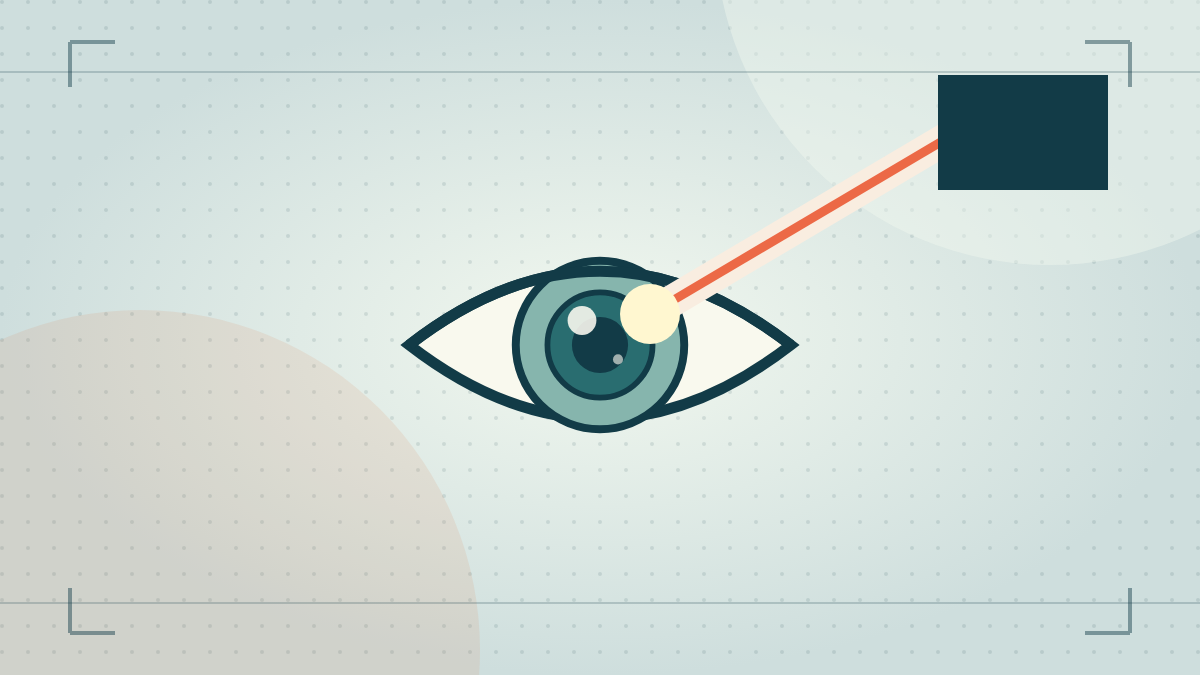
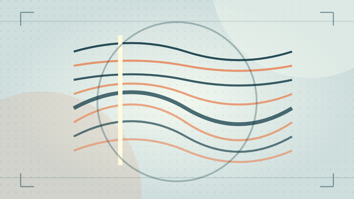
OCT shows retinal layers and can help an eye doctor investigate or monitor some conditions.
A different kind of image
Optical coherence tomography (OCT) uses light to create cross-sectional images of tissue at the back of the eye. Think of it as viewing thin virtual slices through the retina rather than only a flat surface photograph. The test is commonly used alongside an examination, not instead of one.
An eye doctor may use OCT to look for swelling or structural changes associated with conditions such as macular disease. Results need clinical interpretation and context.
What it does not tell you alone
A scan is one part of the picture. Your symptoms, vision measurement, dilated exam, and sometimes other tests still matter. A normal-looking online vision result cannot substitute for retinal imaging when your clinician recommends it.
Common questions
Is OCT the same as a retinal photograph?
No. A conventional photograph shows the retinal surface, while OCT uses light to map cross-sectional layers. An eye doctor may use both because they answer different questions.
Does a scan make the vision chart unnecessary?
No. Imaging shows structure while the chart measures how clearly you identify symbols. Clinicians combine symptoms, examination, visual function, and imaging to interpret a change.
For persistent changes or symptoms, arrange an eye examination. Seek urgent medical care for sudden vision loss, a new curtain or shadow, many new floaters with flashes, severe eye pain, an eye injury, or sudden double vision.
Sources & further reading
This is general educational content based on the linked sources. It is not a diagnosis, prescription, or a substitute for advice from your own eye-care professional. See an error? Contact us.